
Varicose veins affect an estimated 23 percent of adults in the United States, according to the Society for Vascular Surgery. A large portion of those people go years without treatment, assuming the condition is cosmetic. It is not. Varicose veins form when the one-way valves inside leg veins fail, allowing blood to pool and vein walls to bulge outward under increasing pressure. That pressure does not stay contained to the vein itself.
Over time, it damages surrounding tissue, skin, and nerves in ways that become progressively harder to reverse. Research published in the European Journal of Vascular and Endovascular Surgery found that venous disease worsens in approximately 58 percent of untreated patients within five years. The vein specialists at the Foot, Ankle & Leg Vein Center in Boynton Beach treat patients at every stage of this progression.
Chronic Venous Insufficiency Becomes the Foundation of the Problem
Varicose veins are the visible surface signal of a deeper failure called chronic venous insufficiency (CVI). CVI develops when sustained valve dysfunction prevents blood from returning normally from the legs to the heart. The resulting venous hypertension damages capillary walls, allowing red blood cells to leak into surrounding tissue. Hemoglobin from those cells breaks down into hemosiderin, producing the brown skin discoloration characteristic of advanced venous disease.
The CEAP classification system grades venous disease from C0 to C6:
- C0: No visible or palpable signs of venous disease
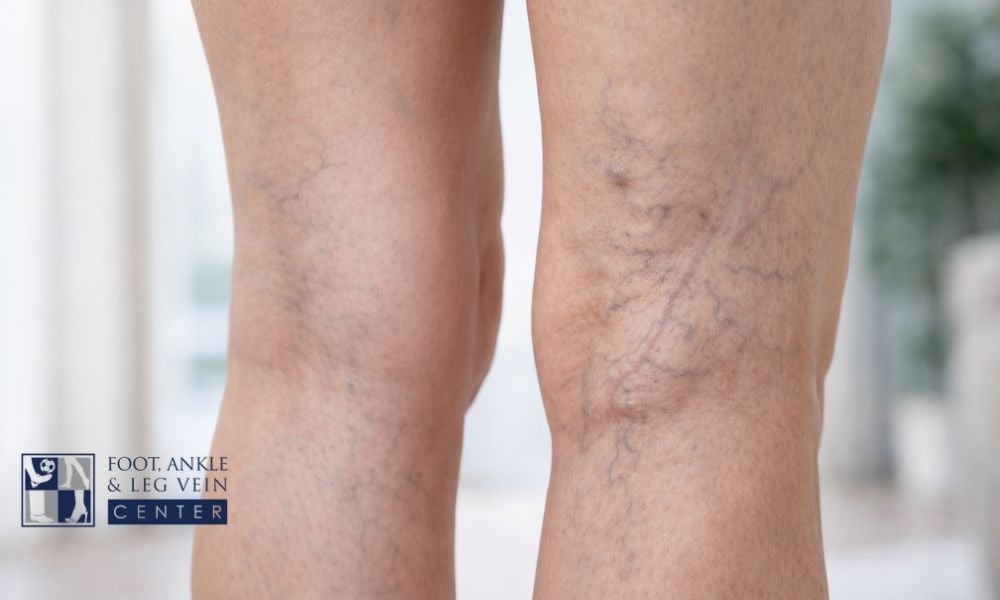
- C1: Spider veins or reticular veins visible at the surface
- C2: Bulging varicose veins present
- C3: Edema present without skin changes
- C4: Skin changes including pigmentation and lipodermatosclerosis
- C5: Healed venous ulcer
- C6: Active open venous ulcer
Dr. Andrew Nicolaides at Imperial College London published research showing that venous hypertension above 80 mmHg is directly associated with the onset of skin changes and ulceration. Patients presenting at C2 can progress to C4 or C5 within three to seven years without any intervention. Swelling, skin thickening, and hyperpigmentation around the lower leg are the early clinical markers of that progression.
Skin Damage and Venous Ulcers Follow Prolonged Pressure
Lipodermatosclerosis is a condition in which the skin and subcutaneous fat beneath the lower leg harden due to chronic venous hypertension. The tissue thickens and scars from the inside, creating a characteristic tapering of the lower leg. This structural change makes the skin fragile. Minor trauma that would heal within days in healthy tissue instead becomes a chronic wound because the underlying circulation cannot deliver enough oxygen and nutrients for normal repair.
Signs that skin damage from venous disease is progressing include:
- Reddish-brown pigmentation spreading above the inner ankle
- Skin surface that feels firm or woody to the touch rather than pliable
- Itching or burning over discolored skin areas
- White, atrophic scars called atrophie blanche where previous skin breakdown healed
- Open sores that fail to close within two weeks despite basic wound care
Venous leg ulcers account for 70 to 80 percent of all chronic leg ulcers, according to the Wound Repair and Regeneration journal. The average venous ulcer takes six months to close and has a recurrence rate above 70 percent without treating the underlying venous insufficiency. The National Library of Medicine reports that venous ulcers are associated with depression, reduced mobility, and lasting quality-of-life impairment. They are largely preventable through early vein treatment.
Blood Clot Risk Increases the Longer Veins Go Untreated
Varicose veins with poor blood flow create the circulatory conditions that favor clot formation. Superficial thrombophlebitis, the development of a clot within a varicose vein close to the skin surface, presents as a firm, red, tender cord along the affected vein. The American Venous Forum estimates this occurs in roughly 125,000 patients annually in the United States. Superficial clots are painful and disruptive, but the greater concern is extension into the deep venous system.
Risk factors that raise the likelihood of clotting in untreated varicose veins include:
- Extended periods of sitting or standing, which further slows venous return
- Long-distance travel without movement breaks
- Dehydration, which increases blood viscosity
- Previous episodes of superficial thrombophlebitis
- Age over 60 combined with limited mobility
A prospective study in the Journal of Vascular Surgery found that patients with untreated varicose veins faced a three-fold higher risk of deep vein thrombosis (DVT) than people without venous disease. A DVT that fragments and travels to the pulmonary vasculature causes pulmonary embolism. The Centers for Disease Control and Prevention estimates that up to 100,000 Americans die from pulmonary embolism each year, making early vein treatment a meaningful preventive step.
Persistent Leg Swelling Leads to Structural Tissue Changes
Early varicose vein disease may produce mild ankle swelling that clears overnight. As venous hypertension worsens over years, fluid leaks from capillaries into the interstitial tissue of the lower leg at a rate the lymphatic system cannot clear. The leaked fluid is rich in proteins and inflammatory mediators that trigger a fibrotic response in surrounding tissue. That tissue begins to scar internally, a process called liposclerosis.
The difference between early and late-stage edema in venous disease is clinically important:
- Early edema: pits when pressed, resolves with leg elevation overnight
- Moderate edema: slower to resolve, leaves indentation marks from socks or shoes
- Late-stage edema: non-pitting, tissue feels firm, does not improve with elevation alone
- Fibrotic tissue: permanently altered structure that compresses local vasculature and nerves
Dr. Hugo Partsch at the University of Vienna demonstrated in a long-term cohort study that patients treated within two years of CVI onset had substantially better outcomes than those who waited five years or more. Once liposclerosis is established, the structural changes are largely irreversible. Compression therapy slows edema but does not reverse fibrosis. The window for preventing permanent tissue change is real and finite.
Nerve Compression and Restless Leg Symptoms Develop Over Time
Chronic venous hypertension does not limit its effects to blood vessels and skin. Elevated pressure in the venous compartment compresses small sensory nerve fibers that run adjacent to the affected veins. Patients with long-standing varicose vein disease frequently describe burning, tingling, or numbness along the lower leg and into the foot. These symptoms mimic peripheral neuropathy but originate from vascular compression rather than primary nerve degeneration.
Neurological symptoms associated with untreated venous insufficiency include:
- Burning or tingling along the inner calf or shin
- Numbness in the foot that worsens after prolonged standing
- Cramping at night that disrupts sleep
- Restless leg sensations including an irresistible urge to move the legs at rest
A 2008 study by Dr. Mark Kahn at the University of San Diego found that 98 percent of patients with confirmed venous insufficiency and restless leg syndrome reported symptom improvement following varicose vein treatment. The finding points to venous pressure as a direct driver of the neurological discomfort. Without treatment, both vascular compression and neurological symptoms intensify over time, adding a layer of chronic impairment on top of the skin and circulatory damage already underway.
What Evaluation Involves and When to Act
A vein specialist uses duplex ultrasound to identify valve reflux, map which veins are failing, and measure the degree of venous hypertension present throughout the leg. This scan is non-invasive and provides real-time venous flow data that physical examination alone cannot deliver. Evaluation is most productive before ulcers, fibrosis, or DVT have developed, when treatment remains straightforward.
Seek evaluation when any of the following are present:
- Bulging veins larger than 3 millimeters visible at the surface
- Leg or ankle swelling that persists into the morning
- Brown discoloration or skin thickening above the ankle
- Aching or leg heaviness that worsens after standing for an hour or more
- Any personal history of clotting in the legs or a previous DVT
Endovenous laser ablation and radiofrequency ablation close failing veins without open surgery, with clinical success rates above 90 percent according to the Journal of Vascular and Interventional Radiology. Both procedures are performed in an outpatient setting with minimal recovery time. Call the Foot, Ankle & Leg Vein Center in Boynton Beach at (561) 725-5066 to schedule a venous evaluation and get a clear picture of where the disease currently stands.